|
IS001 Keynote Lecture: Molecular Mechanisms Underlying Heterogeneities
in Repolarization
Jeanne M. Nerbonne, M.D.
|
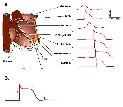 |
| Figure 1. Action potential
waveforms and propagation in the human heart. (A) Schematic
of action potentials, recorded in different regions of the human
heart, are displaced in time to reflect the temporal sequence
of propagation. (B) Schematic of a ventricular action potential
labelled as follows: (0) depolarization; (1) early (fast) repolarization;
(2) plateau phase; (3) late (slow) phase of repolarization;
and, (4) after hyperpolarization/return to the resting membrane
potential. (SA, sino-atrial; AV-atrio-ventricular; RV, right
ventricle; LV, left ventricle) (Journal of Physiology 2000;525(2):285-298). |
|
Copyright © 2000 Japanese
Circulation Society
All Rights Reserved.
webmaster@j-circ.or.jp
|