Japanese Circulation Society
Site mapContact Us
Welcome to JCS web Site
Congres Report
 

AHA/JCS Joint Symposium

 
Vulnerable Plaques: Pathogenesis and Noninvasive Approaches to Detection
 
Plaque Vulnerability and Stabilization: Basic Mechanisms and Molecular Imaging
Masanori Aikawa
Brigham and Women’s Hospital
Harvard Medical School, USA
The Regression of Atherosclerosis: Insights from Mouse Models and Imaging Techniques
Edward A. Fisher
New York University School of Medicine, NY,
NY, USA
An Important Role for Renin Angiotensin System in vulnerable Plaque Formation
Masataka Sata
Department of Cardiovascular Medicine, University of Tokyo
Tokyo, Japan
 
Plaque Vulnerability and Stabilization: Basic Mechanisms and Molecular Imaging
Masanori Aikawa
Brigham and Women’s Hospital
Harvard Medical School, USA
 

Dr. Masanori Aikawa, Brigham and Women’s Hospital, Harvard Medical School, presented data on the role of macrophages in atherogenesis and thrombosis and the effects of lipid-lowering on plaque inflammation and thrombotic complications.

Macrophages are thought to induce thrombotic complications of atherosclerosis through the action of matrix metalloproteinase (MMP) collagenases. Studies in collagenase-resistant and collagenase-deficient mutant mice demonstrated that MMP-collagenases regulate collagen accumulation and organization in mouse atheromata. These studies suggest that MMP-collagenases cause collagen loss in the plaques, leading to plaque vulnerability with subsequent rupture and thrombosis.

Preclinical and clinical studies of lipid-lowering therapy provide further support for the role of inflammation in thrombotic complications of atherosclerosis. Several large statin trials demonstrated a 15% to 40% risk reduction in acute atherosclerotic thrombotic complications without substantially reducing plaque burden.

From 1998 to 2006, Dr. Aikawa and colleagues tested the effects of lipid-lowering on atherosclerotic plaques in rabbit models. The studies demonstrated that lipid-lowering improved a number of features associated with plaque vulnerability and thrombogenicity. Tissue sections of atheromata from cholesterol-fed rabbits showed decreased macrophages after dietary lipid-lowering compared to baseline. Additional histologic studies revealed reduced MMP-collagenase expression and increased collagen accumulation and reduced expression and in situ binding of tissue factor in rabbit atheromata, suggesting potential mechanisms for plaque stabilization. Oxidized LDL (OxLDL) accumulation and endothelial cell activation were decreased from baseline suggesting a potential mechanism for reduced inflammation by lipid-lowering therapy.

A recent study showed that dietary lipid-lowering decreased oxidized phospholipids (OxPL) in rabbit atheromata but increased plasma OxPL in the same rabbits. The mechanisms for this are unclear but OxPL in the blood may serve as a biomarker for plaque stabilization or regression during anti-inflammatory therapy.

Current studies are focusing on molecular imaging of plaque macrophages. One study used 3.0 tesla MRI enhanced with magnetic iron nanoparticles to quantify macrophages in rabbit atheromata. Using this technique, Dr. Aikawa found that cholesterol lowering with statin therapy reduces the magnitude of signal loss in rabbit atheromata. In another study, intravital fluorescence microscopy showed that macrophage accumulation precedes osteogenesis in mouse plaques, suggesting that plaque calcification is related to inflammation. When the mice were treated with statins, macrophage accumulation and osteogenic activity both decreased.

Dr. Aikawa concluded that cholesterol lowering reduces inflammation in atherosclerosis and improves features associated with unstable plaques. Molecular imaging can monitor effects mediated by anti-inflammatory therapies such as lipid-lowering.

 
Page Top
 
The Regression of Atherosclerosis: Insights from Mouse Models and Imaging Techniques
Edward A. Fisher
New York University School of Medicine, NY,
NY, USA
 

An understanding of the mechanisms and factors involved in plaque regression are necessary to limit the risk of coronary artery disease. Dr. Edward A. Fisher, New York University School of Medicine, used mouse models of plaque regression to determine if plaque regression involves a specialized set of factors different from those contributing to plaque progression.

ApoE knock-out (ApoE-/-) mice were fed a high cholesterol diet, increasing their total cholesterol (TC) to >1500 mg/dL. Thoracic segments containing atherosclerotic plaques from these mice were transplanted into wild-type (WT) mice with a normal lipid profile and into control ApoE-/- mice. After 4 weeks, tissue sections of the plaques from the WT mice showed substantial remodeling, with greatly reduced macrophages and development of a fibrous cap. In studies using donors with CD45.1+ foam cells and WT recipients with CD45.2+ macrophages, CD45.1+ cells were found to have migrated from the transplanted plaques to the lymph nodes of the WT recipient mice. Laser capture microdissection and gene expression analysis showed that the phenotype of macrophages from regressing lesions changed to that of migrating dendritic cells, with induction of the chemokine receptor CCR7. Further studies showed that blocking the CCR7 pathway blocked plaque regression. Expression of LXRa, which activates CCR7 gene expression, also was induced in foam cells during regression. LXR agonist treatment turned on expression of CCR7 in plaques of ApoE-/- mice.

Dr. Fisher adapted HDL nanoparticles to carry gadolinium into plaques for MRI and fluorescent dye for microscopic imaging of plaque regression. When injected into ApoE-/- mice, obvious gadolinium uptake and enhancement of the plaques was observed on MRI. In current experiments, Dr. Fisher is incorporating iron oxide and gold particles into the core of HDL particles for use with additional imaging modalities, such as CT imaging. Incorporating copper into the HDL molecule would allow use of PET scanning.

Dr. Fisher concluded that regression of atherosclerosis can be experimentally achieved. In the regression process, foam cells emigrate in a process directed by CCR7 and, in turn, CCR7 is regulated in vivo by LXR. HDL can be modified to improve atherosclerosis imaging, not only by MRI but by multimodal imaging as well. They previously showed that non-enhanced MRI could be used to follow plaque regression/progression in mice, and now the HDL-based agent provides improved capability to non-invasively detect small changes in plaques.

 
Page Top
 
An Important Role for Renin Angiotensin System in vulnerable Plaque Formation
Masataka Sata
Department of Cardiovascular Medicine, University of Tokyo
Tokyo, Japan
 

The renin-angiotensin system plays a critical role in the initiation and progression of atherosclerosis. Dr. Masataka Sata, University of Tokyo, evaluated the role of angiotensin II-angiotensin II type 1 receptor (AT1R) in atherosclerosis progression and destabilization and the role of bone marrow (BM) cells in the atherosclerosis promoting actions of angiotensin II.

The first study evaluated the effects of genetic disruption and pharmacologic blockade of AT1R on plaque progression and instability. Atherosclerotic lesions were significantly reduced by almost 50% in double knock-out ApoE-/- AT1R-/- mice compared with ApoE-/- AT1R+/+ mice (P=0.04). Lipid content was significantly reduced and collagen content significantly increased in the AT1R deficient mice. Pharmacologic blockade of AT1R with telmisartan caused a significant reduction in atherosclerotic lesions compared with mice treated with vehicle or hydralazine. Lipid content was significantly reduced and collagen content was significantly increased in the plaques of telmisartan-treated mice.

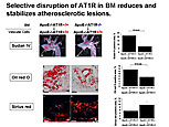
Figure 1. Selective disruption of angiotensin II type 1 receptor (AT1R) in bone marrow reduces and stabilizes atherosclerotic lesions.
【Click to enlarge】
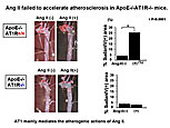
Figure 2. Angiotensin II failed to accelerate atherosclerosis in ApoE-/- AT1R-/- mice.
【Click to enlarge】
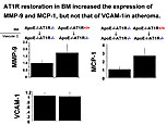
Figure 3. AT1R restoration in bone marrow increased the expression of MMP-9 and MCP-1 but not that of VCAM-1 in atheroma.
【Click to enlarge】

In the second study, angiotensin II infused into hypercholesterolemic mice markedly increased lipid content and decreased collagen content of atheroma compared with vehicle-treated mice (P <0.05). Additionally, accumulation of BM-derived cells in the area of the plaques and BM cell infiltration into plaques increased in mice treated with angiotensin-II.

To investigate the effect of selective AT1R disruption in BM cells, double knock-out chimeric mice with AT1R in vascular cells (ApoE-/- AT1R+/+) but not in BM cells (ApoE-/- AT1R-/-) were generated. These mice had a significant reduction in both atherosclerosis and lipid content of plaques compared to mice with intact AT1R in both vascular and BM cells (Figure 1). Plaque collagen was increased in the chimeric mice but the difference was not significant. Targeted disruption of AT1R in BM cells attenuated accumulation of BM cells in the plaques.

The final study looked at the effect of selective restoration of AT1R in BM of ApoE-/- AT1R-/- mice. Angiotensin-II was infused into ApoE-/- AT1R+/+ and ApoE-/- AT1R-/- mice. After two weeks, angiotensin-II accelerated lesion progression in ApoE-/- AT1R+/+ mice but not in ApoE-/- AT1R-/- mice, suggesting that AT1R mediates the action of angiotensin II (Figure 2). When AT1R was selectively restored in the BM of ApoE-/- AT1R-/- mice, angiotensin-II promoted atherosclerosis and increased lipid and decreased collagen content. Analysis of RNA collected by laser microdissection showed that selective restoration of AT1R in BM increased expression of MMP-9 and MCP-1 but not of VCAM-1 in atheroma (Figure 3).

Dr. Sata concluded that AT1R expressed in BM-derived cells plays a crucial role in the progression and destabilization of atherosclerosis.

 
Page Top