|
|
APSC/JCS Joint Symposium |
| |
|
|
| |
|
| Current Situation of Cardiac Ultrasound in Asia |
| Tsui-Lieh Hsu |
| Division of Cardiology, Taipei Veterans General Hospital |
| Taiwan |
| |
|
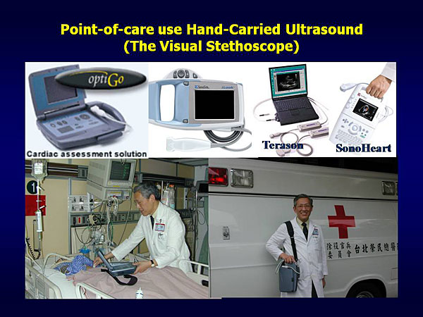
Figure 1. -of-care hand-carried ultrasound.
【Click to enlarge】 |
|
|
|
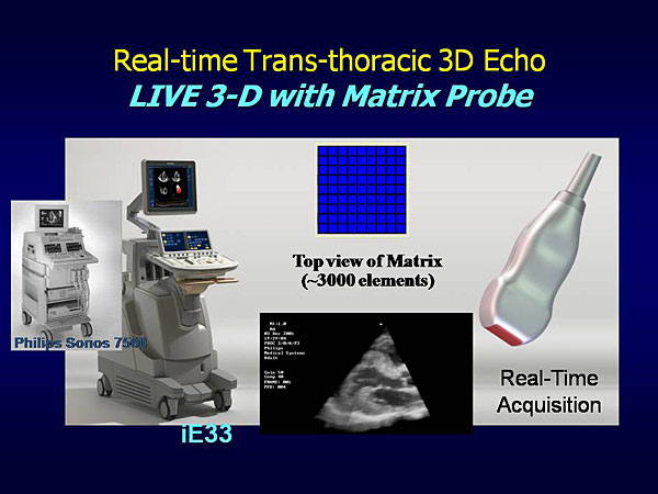
Figure 2. Real-time trans-thoracic 3-D echo.
【Click to enlarge】 |
|
|
The status of cardiac ultrasound in Asia, focusing on advances in technology, training programs, research, professional national echocardiography societies, echocardiography meetings, and future developments, were reviewed by Dr. Tsui-Lieh Hsu, Taipei Veterans General Hospital.
Echocardiography technology has evolved rapidly in the last five decades. The performance and cost of ultrasound systems are important issues. The new generation of ultrasound technology is moving in two directions—toward small, hand-carried devices and toward more comprehensive parametric instruments. Hand-carried and palm-size ultrasound devices cost less and are convenient for extending the physical examination, teaching living anatomy, and using in focused applications (Figure 1). They can also be used for wireless transmission of data to and from remote areas.
Advanced technologies include real-time transthoracic 3-D echocardiography, which uses matrix transducer technology to provide live 3-D imaging (Figure 2). The matrix transducer has been miniaturized for use in the “real-time 3-D TEE,” which is useful in the perioperative setting for decision-making.
Laboratories in most Asian countries are not using the latest technology because of the cost. However, costs are decreasing and future goals are to have entirely digital echocardiography laboratories with matrix transducer technology for full spectrum diagnostic applications, using quantitative analysis software. Such a system can be used for data management, archiving, retrieving, and transferring data. Computer-assisted devices and other peripheral parts can also be used in conjunction with the system.
Most Asian echocardiography training programs are based on the ASE/ACC/AHA recommendations. The various echocardiography modalities are used in basic research to clinical applications throughout Asia. Cardiac ultrasound research is very productive in Japan, Korea, Hong Kong, and Australia. Most Asian countries have national echocardiography societies that train physicians, disseminate information, share skills, publish research, hold annual meetings, and develop guidelines and consensus statements. For example, the Japanese Society of Echocardiography (JSE) was founded in 1990 and currently has 2,000 members. JSE has published the Journal of Echocardiography since 2003. Many regional echocardiography meetings are held in Asia, including the World Congress of Echocardiography and Vascular Ultrasound (ISCU), International Cardiac Doppler Society (ICDS), and the Asia-Pacific Cardiac Doppler Echocardiography (APCDE) meetings.
Future developments will include establishment of a partnership program between the national echocardiography societies in Asia, concluded Dr. Hsu. An Asia-Pacific working group of echocardiography professionals for communication and sharing of resources needs to be developed. He also suggested founding an Asia-Pacific Society of Echocardiography for promoting education, research, and clinical work in the field of cardiac ultrasound.
|
| |
| Page Top |
|
| |
|
| Nuclear Cardiology in Asia: Current Status |
| Kevin C. Allman, MD |
| Royal Prince Alfred Hospital |
| Sydney, Australia |
| |
|
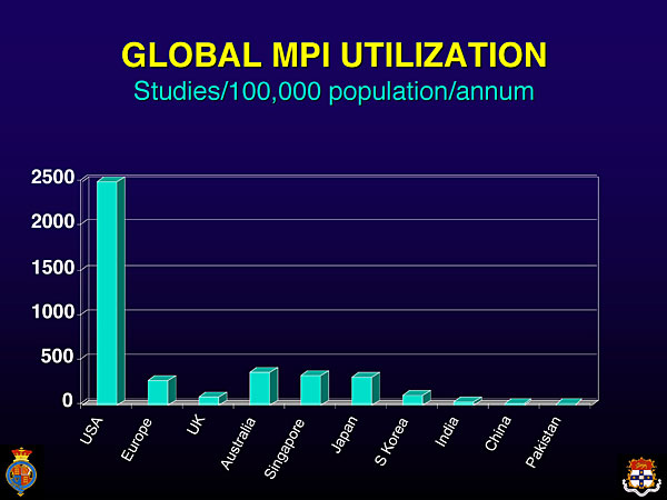
Figure 1. Global use of myocardial perfusion imaging.
【Click to enlarge】 |
|
|
|
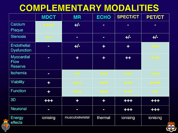
Figure 2. Imaging modalities that are complementary.
【Click to enlarge】 |
|
|
The clinical and research aspects of nuclear cardiology in Asia were reviewed by Dr. Kevin C. Allman in this lecture. Currently used imaging modalities in nuclear cardiology are single photon emission-computed tomography (SPECT) and positron emission tomography (PET).
SPECT is widely validated and used in Asia and the rest of the world for assessing myocardial perfusion and function. The largest user of SPECT is the United States, which performs 7 million studies annually. Utilization is much lower in the Asia-Pacific region partly because of restricted access (Figure 1).
A chart comparing the different imaging modalities used in cardiology shows that SPECT is just as good as magnetic resonance (MR) and echocardiography for evaluating cardiac ischemia, viability, and function (Figure 2). Quantitation is better with PET, allowing assessment of earlier phases of disease, including myocardial flow reserve and endothelial dysfunction. Nuclear cardiology techniques are better than other modalities for assessing cardiac neuronal function.
To diagnosis coronary artery disease (CAD):
- Computed tomographic angiography (CTA) detects earlier disease before stenosis with a very high negative predictive value (NPV).
- SPECT is comparable to echocardiography and MR.
To evaluate the extent of CAD:
- PET is better than SPECT for multivessel disease.
- SPECT and echocardiography are similar.
- Data are limited for PET and MR.
To assess viability:
- SPECT, PET, MR, and echocardiography are comparable.
- MR provides images of transmurality of MI versus SPECT.
To assess function:
- SPECT, echocardiography, MR, and PET all perform well.
Tracers are used to expand the usefulness of imaging techniques. For example, the norepinephrine analog, metaiodobenzylguanidine (MIBG) is useful for assessing cardiac neuronal function, heart failure prognosis, arrhythmias, cardiomyopathy, and high-risk patients for defibrillator implantation. I-123 BMIPP fatty acid, referred to as ischemic memory, can be used to detect areas of prior myocardial ischemia, emergency assessment of chest pain, and prognostic assessment in patients with CAD.
Future nuclear technologies include new types of gamma cameras that provide greater sensitivity for SPECT imaging, fast SPECT cameras, dynamic SPECT to measure blood flow (MVD), office-based dedicated PET cameras, PET quantitation software, lower radiation doses using PET tracers and new SPECT scanners, hybrid imaging (i.e., CTA followed by MPI), cheaper generic tracers, and gated SPECT to assess LV dyssynchrony.
Cardiologists in Asia need to prepare for appropriate use of new technologies, concluded Dr. Allman. Increased demand for clinical studies will require strategies for instrumentation and an adequate workforce of technologists and nuclear cardiologists. Professional societies have an important role in planning and education to deal with these issues. |
| |
| Page Top |
|
| |
|
| Prudent Use of New Coronary Imaging Methods in the Real World |
| Kui Hian Sim |
| Sarawak General Hospital and University Malaysia Sarawak, Malaysia |
| |
Cardiovascular disease is rapidly becoming the number one killer in Asia, resulting in burdens of death and disability that outweigh those imposed by communicable diseases. The scope of the problem is huge, with a population of 3,415,000 people in the Asia-Pacific region, who live in widely varying socio-economic conditions. Dr. Kui Hian Sim, University Malaysia Sarawak, discussed the challenges and issues of cardiovascular disease management in the region.
|
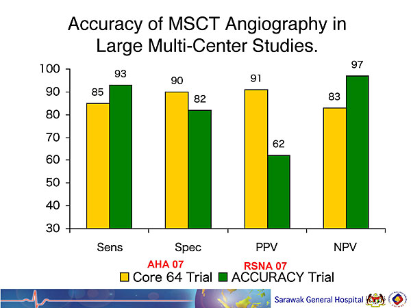
Figure 1. Accuracy of multi-slice computed tomography (MSCT) angiography in large multi-center studies.
【Click to enlarge】 |
|
|
|
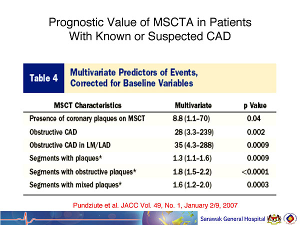
Figure 2. Prognostic value of MSCT angiography in patients with known or suspected CAD. (Reprinted from JACC, 49(1), Pundziute G, Schuijf JD, Jukema JW, Boersma E, de Roos A, van der Wall EE, Bax JJ, Prognostic value of multislice computed tomography coronary angiography in patients with known or suspected coronary artery disease,62-70., 2007, with permission from Elsevier.)
【Click to enlarge】 |
|
|
|
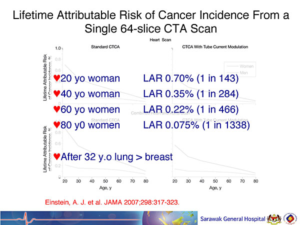
Figure 3. Lifetime attributable risk of cancer incidence from a single 64-slide CTA scan. (Reprinted from JAMA, 298(3), Einstein AJ, Henzlova MJ, Rajagopalan S, Estimating risk of cancer associated with radiation exposure from 64-slice computed tomography coronary angiography, 317-323, 2007, with permission from American Medical Association.)
【Click to enlarge】 |
|
|
Challenges for cardiologists in managing the epidemic of coronary disease in Asia include availability, accessibility, affordability, and appropriate utilization of healthcare technologies. Medical litigation can also be an issue. Diagnostic coronary imaging is the most important aspect of cardiovascular disease management. Important imaging modalities include echocardiography, angiography, cardiac magenetic resonance imaging (MRI), computed tomography (CT), and nuclear imaging techniques such as single photon emission-computed tomography (SPECT) and positron emission tomography (PET). These and other tests are used to assess morphology and function of the myocardium, cardiac valves, coronary arteries, and ventricles, and to evaluate ischemia and myocardial viability.
Cardiac CT is a popular imaging modality in Asia, with a total of 700 CT scanners installed in the region. In the last 3 years, 230 64-slide multi-slice CT (MSCT) scanners have been acquired. CT allows effective specific therapeutic intervention and improves patient outcomes. The major costs and complications associated with invasive coronary angiography often can be avoided by using CT angiography. In the CORE 64 and ACCURACY trials, MSCT sensitivity was 85% and 93%, specificity 90% and 82%, PPV 91% and 62%, and NPV 83% and 97%, respectively (Figure 1). A study of the prognostic value of MSCTA in patients with known or suspected coronary artery disease (CAD) reported a 0% first-year event rate in patients with normal arteries on MSCT and a 63% first-year event rate in patients with any evidence of CAD on MSCT. Multivariate predictors of events in this study are shown in Figure 2. Another study demonstrated that MSCTA results have prognostic value for prediction of all-cause mortality (risk-adjusted P <0.001).
The benefits of CT cardiac imaging need to be weighed against the risks of radiation exposure. Several studies have demonstrated increased risk of cancer with CT. One study found that 1 of every 143 women who are scanned once at age 20 will get cancer, usually breast, compared to 1 in 3,261 in the general population (Figure 3). Dr. Sim concluded that pre-disease screening offers opportunities and impacts disease management. CT and MRI are emerging technologies. Collaboration is needed to gather data on cardiovascular imaging modalities in the Asia-Pacific region. |
| |
| Page Top |
|
| |
|
|