Japanese Circulation Society
Site mapContact Us
Welcome to JCS web Site
Congres Report
 

KSC-JCS Joint Symposium

 
Novel Therapeutic Strategies against Heart Failure
 
Novel Therapeutic Strategies Against Heart Failure on the Molecular Mechanisms and New Therapeutic Targets.
Eun-Seok Jeon, MD, PhD

Division of Cardiology, Samsung Medical Center, Korea

Identification of Target Domains of the Cardiac Ryanodine Receptor to Correct Channel Disorder in Failing Hearts.
Takeshi Yamamoto
Department of Medicine and Clinical Science, Yamaguchi University Graduate School of Medicine, Ube, Japan
p53 as a New Therapeutic Target for Heart Failure.
Tohru Minamino
Department of Cardiovascular Science and Medicine, Chiba University Graduate School of Medicine, Japan
 
Novel Therapeutic Strategies Against Heart Failure on the Molecular Mechanisms and New Therapeutic Targets.
Eun-Seok Jeon, MD, PhD

Division of Cardiology, Samsung Medical Center, Korea


Cardiac remodeling is influenced by six molecular pathways in addition to hemodynamic load and neurohormonal activation. These pathways provide potential therapeutic targets for heart failure (HF). In this presentation, Dr. Eun-Seok Jeon, Samsung Medical Center, Korea, reviewed studies of these molecular pathways and targets.

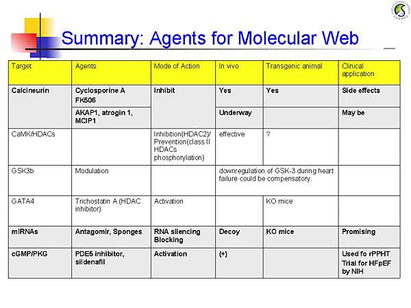
Calcineurin dephosphorylates NFAT, which translocates to the cell nucleus and activates genes that cause hypertrophy. Endogenous calcineurin inhibitors–AKAP1, atrogin 1 (FBXO32), and RCAN1– are being studied as inhibitors of this pathway. Calmodulin and its kinase contribute to cardiac hypertrophy through HDAC and RyR phosphorylation.

Glycogen synthase kinase 3b (GSK3b) normally inhibits hypertrophy but is suppressed by Akt or PKA phosphorylation, allowing progression of hypertrophic pathways. Experiments in mouse models showed that persistent GSK3b inhibition induces compensatory hypertrophy, inhibits apoptosis and fibrosis, and increases cardiac contractility.

Deletion of the transcription factor GATA4 in mouse models results in progressive cardiac dysfunction and dilation, with pressure overload causing rapid decompensatory HF. GATA4 regulates cardiac gene expression, hypertrophy, stress compensation, and myocyte viability.

MicroRNAs (miRNAs) mediate post-transcriptional gene silencing. In vitro overexpression of miRNA-133 inhibited cardiac hypertrophy. In vivo miRNA-133 inhibition caused marked, sustained hypertrophy. MicroRNA-208-/- mice had reduced hypertrophy in response to pressure overload. These data show that these miRNAs are key regulators of cardiac hypertrophy.

Cyclic GMP (cGMP)-dependent protein kinases regulate phosphodiesterases (PDE). The PDE5A inhibitor sildenafil suppresses hypertrophy and improves heart function in mice with induced pressure overload and reverses pressure overload-induced hypertrophy. An ongoing NIH-sponsored trial is testing PDE5 inhibitor therapy for HF with preserved EF.

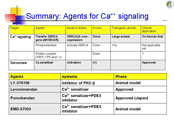
Ca2+ is the central regulator of excitation-contraction coupling. Potential therapies in calcium signaling pathways include SERCA2A gene transfer, phospholambam gene deletion, and production of constitutively phosphorylated phospholambam, all of which improved heart function and remodeling in animal models. A phase 1/2 trial is investigating AAV1/SERCA2a gene transfer in patients with NYHA III/IV HF with EF ≤30%. The initial outcomes of this trial will provide valuable data for molecular targeted therapy for HF.


Figure 1. Agents for the molecular web
【Click to enlarge】
Figure 2. Agents for calcium signaling.
【Click to enlarge】

Calcium sensitizers stimulate cardiac contractility without causing intracellular calcium overload or increasing myocardial oxygen demand. Mutations in the titin cap (T-cap) gene that promote association of T-cap with titin and calsarcin 1 cause hypertrophic cardiomyopathy. Mutations that impair this association cause dilated cardiomyopathy. Manipulating these interactions might provide a new therapeutic approach for HF.

Investigation of novel agents targeting molecular pathways (Figure 1) and calcium signaling pathways (Figure 2) is promising for the treatment of HF.

 
Page Top
 
Identification of Target Domains of the Cardiac Ryanodine Receptor to Correct Channel Disorder in Failing Hearts.
Takeshi Yamamoto
Department of Medicine and Clinical Science, Yamaguchi University Graduate School of Medicine, Ube, Japan
 

The ryanodine receptor (RyR) is a calcium release channel on the sarcoplasmic reticulum (SR). Mutations in either the N-terminal or central domain of RyR weaken interactions between the two domains, destabilizing the channel. Dr. Takeshi Yamomoto, Yamaguchi University Graduate School of Medicine, previously reported that a novel 1,4-benzothiazepine derivative K201 (JTV519) stabilizes the RyR by zipping the N-terminal and central domains. The aim of this study was to identify the mechanism by which K201 stabilizes the RyR channel.

A synthetic peptide was made that corresponds to the RyR2 sequence 2114-2149 (DP2114-2149) and is similar to annexin V, which binds RyR2. Quartz Crystal Microbalance (QCM) was used to test K201 binding to the synthetic peptide. QCM showed that K201 binds to the expressed RyR2 fragments 1741-2270 and 1981-2520, both of which contain the domain 2114-2149. A second experiment showed that K201 binds to purified RyR2.

Further experiments showed that, like K201, DP2114-2149 inhibits FK506-induced calcium leakage from RyR2. Dp2114-2149 also inhibited FK506-induced calcium spark in normal cardiomyocytes (p<0.01) and a spontaneous calcium spark in failing cardiomyocytes (p<0.01).

Figure 1. Zipping-unzipping conformation visualized by quencher accessibility.
【Click to enlarge】

The channel stabilization mechanism of DP2114-2149 was investigated using specific fluorescence labeling of the RyR2 and visualizing the zipping-unzipping conformation by fluorescent quencher accessibility. In the failing SR, the N-terminal and central domains were unzipped at baseline; addition of Dp2114-2149 zipped the two domains (Figure 1).

Next, the Dp2114 binding domain of RyR2 was fluorescence labeled. The conformation of domain 2114-2149 and its counterpart were analyzed by fluorescent probe. When the N-terminal and central domain pair zipped, DP2114 and its binding size unzipped, and vice versa. In the failing SR, domain 2114-2149 and its counterpart were unzipped by K201.

Figure 2. The current model.
【Click to enlarge】

The current model based on these experiments shows that in the destabilized channel, the N-terminal and central domain are unzipped, while domain 2114-2149 and its counterpart are zipped (Figure 2). Addition of K201 or DP2114-2149 stabilizes the channel by unzipping domain 2114-2149 and its counterpart, causing the N-terminal and central domains to zip.

In summary, K201 binds to domain 2114-2149 of RyR2. DP2114-2149 has a strong stabilizing effect on RyR2, similar to K201. K201 unzipping of domain 2114-2149 and its counter domain causes zipping of the N-terminal and central domain pair, stabilizing RyR2.

Dr. Yamamoto concluded that domain 2114-2149 and its counter domain play an important role in stabilizing RyR2. These domains might be a novel target for HF treatment.

 
Page Top
 
p53 as a New Therapeutic Target for Heart Failure.
Tohru Minamino
Department of Cardiovascular Science and Medicine, Chiba University Graduate School of Medicine, Japan
 

Dr. Tohru Minamino, Chiba University Graduate School of Medicine, presented a series of experiments investigating the role of p53 in heart failure (HF). Dr. Minamino hypothesized that cardiac angiogenesis might play an important role in the development of cardiac hypertrophy and prevent HF. This hypothesis was tested in a pressure overload mouse model using thoracic aortic constriction (TAC).

In this mouse model, the heart to body weight ratio and left ventricular (LV) wall thickness increased, peaking on day 14 after TAC (adaptive phase). After day 14, the heart/body weight ratio decreased (maladaptive phase). Systolic function was preserved at day 14 but impaired by day 28. In the adaptive phase, capillary density increased but decreased during the maladaptive phase. Angiogenic factor expression was upregulated during the adaptive phase and downregulated during the maladaptive phase. Treatment with the angiogenesis inhibitor TNP-470 caused decreased LV wall thickness and impaired systolic function. Promotion of angiogenesis with angiogenic factors enhanced LV wall thickness and preserved systolic function under sustained pressure overload.

HIF-1 was upregulated in the adaptive phase and downregulated in the maladaptive phase. In cardiomyocyte-specific HIF-1a knockout mice, VEGF expression and angiogenesis were decreased and systolic function was impaired. Histologic analysis showed that cardiac ischemia began 3 days after TAC and was sustained through day 28. Sustained hypoxia induces p53 expression, thereby inhibiting HIF-1 activity. Analysis showed that p53 was markedly upregulated in the adaptive phase, which was associated with HIF-1 and VEGF downregulation.

Figure 1. Preserved cardiac function in p53 KO mice
【Click to enlarge】

In p53 knockout mice, 28 days after TAC, angiogenesis and hypertrophy were enhanced and systolic function was improved (Figure 1). Activation of p53 with quinacrine decreased angiogenesis and impaired systolic function. Inhibition of angiogenesis by p53 caused endothelial cell death followed by cardiomyocyte death. These results show that HIF-1 dependent angiogenesis is crucial for the adaptive response. However, ischemia induces p53 expression, which inhibits angiogenesis, leading to HF.

Using expression cloning, STUB1 (CHIP) was identified as an endogenous suppressor of p53 in the heart. Experiments showed that STUB1, a chaperon-associated ubiquitin ligase, negatively regulates p53 activity by ubiquitination. STUB1 overexpression was downregulated after MI, leading to a marked increase of p53. STUB1 overexpression attenuated p53 accumulation, improving systolic function and preventing cardiac remodeling after MI.

These studies demonstrate the critical role of p53 in HF. Further investigation of p53 and cellular aging signals in the heart will provide new insights into the treatment of HF.

 
Page Top