Japanese Circulation Society
Scientific Sessions Activities Publications
index
>congress report>the64th scientific session>invited lecture
Invited Lecture

Pathophysiology of the Atheroma and Mechanisms of Plaque Rupture
Peter Libby, M.D.
Brigham & Women's Hospital
Harvard Medical School
Boston, MA, USA
  • Time course of human atherogenesis
  • Molecular mediators of the atherosclerotic process
  • Complications of atherosclerosis
  • Biologic effects of lipid lowering therapy
  • Closing

  • Atherosclerosis was originally thought of as rust encrusting the lumen of the artery. However, it is now known that the normal artery wall is comprised of arterial smooth muscle cells and endothelial cells, and when a nascent atherosclerotic plaque is present the artery wall becomes a host to T lymphocytes and macrophages. The current understanding of the pathophysiology of the atheroma, the molecular mechanisms of plaque rupture, as well as the mechanisms likely responsible for the effects of lipid lowering therapy were reviewed in this lecture.

    PAGE TOP


    Time course of human atherogenesis


    Figure 1. Schematic time course of human atherogenesis (Libby 2000)
    Click to enlarge


    The infiltration of macrophages is one hallmark of the atherosclerotic lesion (Fig. 1). The arrival of leukocytes in the artery wall is one of the first changes when an atherogenic diet is initiated, as shown by work by Ross. He showed leukocyte adherence to an intact endothelial mono layer early in the atherosclerotic process in the aorta of a non-human primate fed a diet rich in cholesterol and fat. Transmission electromicroscopy shows how intimately the membrane of the monocytes interdigitate with the endothelial cell.

    Pioneering observations by Poole and Florey in the 1950s showed that normal endothelial cells resist prolonged contact with endothelial cells by a variety of mechanisms, including the ability to make nitric oxide (NO) constitutively. NO can inhibit the adhesion of leukocytes to the endothelial cell. Endothelial nitric oxide synthase (NOS) is regulated by shear stress. So, in areas of normal laminar shear there are high levels of NO relative to areas in the circulation without normal shear, such as flow dividers and branch points, where increased leukocyte adhesivity can occur in part due to the decrease in NO.

    PAGE TOP


    Molecular mediators of the atherosclerotic process


    The underlying molecular mechanisms associated with the large number of macrophages present in the atheroma are beginning to be appreciated. Mononuclear cells are recruited into the artery wall by leukocytes attached to the endothelium. This leukocyte attachment is mediated by various leukocyte adhesion molecules expressed on the surface of the endothelial cell, which engage counter receptors, or cognate ligands, on various classes of leukocytes.

    A combination of careful observation and traditional biochemical, morphologic and cell biologic work has led to the definition of candidates for the various ways in which macrophages are recruited and activated during atherogenesis. Work with genetically-engineered animals has yielded information that 1) implicates vascular cell adhesion molecule-1 (VCAM-1) in the adhesion reaction, among other adhesion molecules, 2) shows monocyte chemoattractant protein-1 (MCP-1) is a chemoattractant causing the directed migration of leukocytes, and 3) shows macrophage colony stimulating factor (M-CSF) is a co-mitogen and activator that can cause the expression of scavenger receptors on macrophages. Libby reviewed work performed in his laboratory that provides this new understanding.


    Figure 2. VCAM-1 expression in rabbit aorta (Arteriosclerosis, Thrombosis, Vascular Biology 1993;13:197-204.)
    Click to enlarge
    Reprinted with permission from Lippincott Williams and Wilkins (www.lww.com).

    VCAM-1 and leukocyte adhesion

    Vascular cell adhesion molecule-1 (VCAM-1) is an adhesion molecule of particular interest in the context of atherosclerosis because it finds only those monocytes and T-cells present in the nascent atherosclerotic lesion. VCAM-1 is expressed by the endothelium of nascent fatty streaks and by microvessels in the mature human atherosclerotic plaque. VCAM-1 is a putative molecular mediator of the leukocyte adhesion reaction, as shown by Li in work in Libby's laboratory. In rabbits fed a cholesterol-rich diet she showed that VCAM-1 is not expressed on normal rabbit endothelium, but there was increased expression of VCAM-1 by week one of the diet (Fig. 2). At three weeks, VCAM-positive endothelial cells with adherent leukocytes were present.

    MCP-1 mediates leukocyte penetration

    One candidate mediator for leukocyte penetration into the nascent atheroma studied was a potent monocyte chemoattractant protein (MCP-1) produced by endothelial and smooth muscle cells and localized in human and experimental atherosclerotic plaque. However, proving that a molecule is functioning as a mediator is difficult. Genetically engineered mice are now being used as models to test causality and prove whether or not a particular candidate molecule is important.


    Figure 3. Reduced lipid deposition in MCP-1 deficient atherosclerotic mice (Molecular Cell 1998;2:275-281.)
    Click to enlarge

    The absence of MCP-1 in a genetically-engineered hyperlipoproteinemic mouse plays an important role in atherogenesis, as shown by work by Libby's group in collaboration with Rollins. They found accumulation of lipid in the arch and the distal aorta in an LDL-receptor defective animal competent to make MCP-1 and in the MCP-1 wild type animal. But, there was a marked reduction in lipid lesions in the double-knockout animal lacking LDL receptors and the gene to encode MCP-1 (Fig. 3). MCP-1 plays a causal role in the recruitment of leukocytes into the mouse atheroma. But, once the monocytes have inhabited the atherosclerotic plaque, the monocytes are activated and change phenotype, becoming a macrophage that expresses scavenger receptors and engulfs modified lipoproteins to become a foam cell that can divide. The division of monocyte macrophages and the division of the smooth muscle cells in the advanced human atherosclerotic plaque and in the experimental atheroma in rabbits are equally as common.


    Figure 4. M-CSF deficiency reduces atheroma in LDL receptor knockout mice (J Clinical Investigation 1998;101:2702-2710.)
    Click to enlarge
    Republished with permission of Journal of Clinical Investigation from Heterozygous osteoporotic (op) mutation reduces atherosclerosis in LDL receptor-deficient mice, Rajavashisth, 101(12), 1998; permission conveyed through Copyright Clearance Center, Inc.

    M-CSF activates leukocytes

    Although it is clear that the leukocytes become activated during atherosclerosis, the molecular mediator of this process is unknown. Work from the 1980s and 1990s provides some candidates. Libby's laboratory has had a long interest in macrophage colony stimulator factor, (M-CSF) a potent monocyte activator and co-mitogen produced by endothelial and smooth muscle cells and localized in human and experimental atheroma. Recently they were able to prove causality of M-CSF in macrophage function in atherosclerosis using a double knockout osteopetrotic mouse. Two features that distinguish their approach are the homogeneous C57Bl6 background of the mice and the defined diet that provided an adequate diet without weight loss. In the M-CSF-wild type mice there was a great deal of lesion formation. In the one allele-M-CSF-gene defective mice there was marked diminution in the fatty lesions, while atherogenesis was nearly absent in the M-CSF-gene defective mice with no functional M-CSF (Fig. 4). Thus, they were able to define a gene dosage-related effect for M-CSF in the generation of lesions in this experimental model of atherosclerosis.

    PAGE TOP


    Complications of atherosclerosis


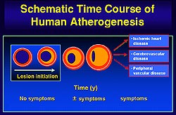
    The presence of the mature atherosclerotic lesion sets the stage for complications of atherosclerosis. Patients may present with symptoms from stenotic lesions or with thrombotic symptoms, such as acute myocardial infarction or unstable angina, which may occur without warning as in chronic stable syndromes of atherosclerosis.

    Much has been learned about the pathogenesis of the thrombotic complications of atherosclerosis. 
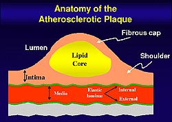
The fibrous cap of the plaque protects the integrity of the atheroma, providing a shield between the thromogenic material in the plaque's lipid core and the coagulation factors present in the circulating blood. Weakening of the fibrous cap leads to plaque rupture. The thrombogenicity of the plaque's lipid core also plays a key role in the unstable coronary syndrome.

    Libby reviewed some of the thinking and experimental results advocated by his laboratory that helps to explain the features of the unstable atheroma. When inflammation is present in the intima the leukocyte can send signals to the smooth muscle cell to inhibit the biosynthesis of collagen, which is the key structural component of the plaque's fibrous cap. The leukocytes can also exchange signals that cause overexpression of collagen-degrading proteinases. Thus, inflammation places the collagen in the plaque's fibrous cap under the double attack of decreased synthesis and increased breakdown, setting the stage for plaque rupture and thrombosis. The leukocytes can also exchange signals that can augment the production of the procoagulants that make it dangerous for the blood to enter the artery wall.

    In an experiment with human smooth muscle cells in culture to measure collagen biosynthesis it was shown that the smooth muscle cells incorporate proline, an amino acid rich in collagen, in the basal state into newly synthesized collagen. When the smooth muscle cells are exposed to platelet-derived growth factor (PDGF) or transforming growth factor-beta, which are released during coagulation, there was an increase in the biosynthetic rate of collagen. For lesion healing this is very important. But, when smooth muscle cells are exposed to gamma interferon, a T-cell derived cytokine, new collagen synthesis by smooth muscle cells was nearly inhibited. It is now recognized that there are many T-cells in various regions of the plaque, particularly those prone to rupture.

    Role of gamma interferon

    Van der Wal observed some years ago that in human fatal thrombotic events in coronary arteries, macrophages and T-lymphocytes are the dominant cell types whether or not there is thrombosis due to plaque rupture or superficial erosion. Further, the cells in that region overexpressed HLA-DR, a transplantation antigen. Libby's laboratory defined that antigen more than a decade ago as a gamma interferon-inducible structure on the surface of the smooth muscle cell. So, the recent clinical finding that there are HLA-DR-positive smooth muscle cells at the sites where plaque ruptures is strong evidence of gamma interferon action at the site of rupture of human atheroma. Gamma interferon can weaken the fibrous cap by inhibiting biosynthesis of new collagen by the smooth muscle cell, impairing the ability of the smooth muscle cell to repair and maintain the plaque's fibrous cap. The level of collagen in the fibrous cap depends on the rate of synthesis as well as the rate of breakdown. The triple collagen fiber is ordinarily a very strong, biochemically-resistant structure.

    Role of matrix metalloproteinase

    Only a handful of enzymes are capable of attacking collagen, notably the interstitial collagenases. The interstitial collagenases, members of the matrix metalloproteinase (MMP) family, can make an initial proteolytic cleavage, breaking the collagen fiber into three-quarter and one-quarter fragments. Normal human arteries fortunately do not contain active forms of interstitial collagenase.

    Libby's group showed some years ago that foam cells derived from both macrophages and smooth muscle cells in the mature human atherosclerotic plaque overexpress the collagen- degrading enzyme MMP-1. However, the presence of immunoreactive MMPs do not imply biologic activity. MMPs are synthesized as pro-enzymes that require processing to attain activity. Antibodies do not distinguish the zymogen from the active form of the MMP. Potent endogenous inhibitors of the matrix MMP are widely distributed. Libby's laboratory has described three of the four known tissue inhibitors of MMP found in the atheroma.

    Recent work by Libby's laboratory shows there are two enzymes, MMP-1 and MMP-13, expressed in the atherosclerotic plaque that can break down collagen, including in situ evidence of collagenolysis by MMP in the human atherosclerotic plaque. However, a break in the fibrous cap would not matter but for the ensuing thrombosis.

    What causes the thrombogenicity of the lipid core?

    For more than a decade it has been known that macrophages in the core of the human atherosclerotic plaque overexpress tissue factor, a potent procoagulant. But, until recently the molecular switch that turns on tissue factor expression was unknown. The usual soluble cytokines localized in the atheroma do not induce macrophage tissue factor expression. T cells can induce macrophage tissue factor by contact, but the signal remained elusive.

    Recent work from Libby's laboratory likely identifies the missing link between the T cell and the lymphocyte, which are side by side when the human atherosclerotic plaque ruptures. They have studied a new signaling system known as CD 40 ligand, a surface bound signal. The usual soluble cytokines do not induce appreciable tissue factor activity, but recombinant CD 40 ligand and the membranes of activated T cells in a CD-ligand dependent manner briskly induce tissue factor gene expression. The target gene product tissue factor is expressed in the same cell that gives rise to the receptor CD 40 that binds CD 40 ligand. Thus, they believe that CD 40 turns on tissue factor expression during an inflammatory state in the arterial intima.

    PAGE TOP


    Biologic effects of lipid lowering therapy


    Lipid lowering therapy can reduce clinical events. Libby reviewed data suggesting that lipid lowering may be affecting the very biology of the atherosclerotic process and improving it in a manner that can stabilize the human atherosclerotic lesion.

    In the rabbit model atheroma was created by a combination of balloon injury and a high cholesterol diet. Some animals then remained on the high cholesterol diet for 16 months, a period of time similar to that needed to accrue clinical benefit from statins in the mega trials. Other animals were shifted to a low cholesterol diet during which time their cholesterolemia gradually declined towards normal for a rabbit. The baseline lesions in the rabbits were similar to human atherosclerotic plaque lesions created in this manner. The lipid core was filled with macrophages overexpressing the collagenolytic enzyme MMP-1.

    With the continued high cholesterol diet, many macrophages remained and expressed collagenase. In the animals switched to the low cholesterol diet there was a marked decrease in the number of macrophages and in the amount of collagen-degrading matrix MMP-1. Thus, the plaque in the low cholesterol rabbits has a more complex, reinforced collagenous skeleton, compared to the filamentous, scant network of collagen in the intima of the animals continued on a high cholesterol diet. In this same model Aikawa also showed that tissue factor protein overexpression by the macrophages in the baseline lesion persists with high cholesterol feeding, but declines strikingly with the low cholesterol diet. The activity of tissue factor protein can be gauged by its ability to bind Factor VIIa and Factor Xa.

    This series of experimental observations suggest that in rabbits with diet-induced atherosclerosis, reduced cholesterol consumption could limit inflammation, improve features of plaque associated with stability in humans, and reduce thrombogenicity. This provides a potential mechanistic explanation for the striking benefit of lipid lowering therapy.

    PAGE TOP


    Closing


    Figure 5. Anatomy of the atherosclerotic plaque (Libby 2000)
    Click to enlarge

    In years past, the focus at a cardiology conference would have been the heart muscle itself. Now it is recognized that the blood vessel is the major culprit and the heart muscle is an innocent bystander in the most important cardiovascular diseases.

    Atherosclerosis begins when there is an excess of risk factors (Fig. 5). Presently, the most understood risk factor is LDL. Excess LDL in the artery wall can become modified and instigate an inflammatory response on the endothelial surface of the artery. This causes expression of leukocyte adhesion molecules like VCAM-1 that can bind to cognate receptors. MCP-1 can then cause diapedesis of the cells into the artery wall and M-CSF can augment the expression of scavenger receptors. This allows the macrophages to engulf the modified lipoprotein particles and transform them into foam cells, the hallmark of the fatty streak and the initial lesion of atherosclerosis that initially grows outward.

    Lesion complication begins when the smooth muscle cells in the intima divide. Other smooth muscle cells in the tunica media migrate into the intima where they divide and lay down collagen to make a fibrofatty lesion, an intermediate form of the atherosclerotic lesion. Persistence of risk factors such as high levels of LDL over a period of time provides the opportunity for the lipid core to grow, usually in an outward direction thus preserving the lumen. The inflammatory response smolders. The T cells send soluble signals to the macrophage or can activate the macrophage by contact through CD 40 ligand. The activated macrophage in turn secretes many mediators, including the matrix MMPs which can degrade the collagen that lends strength to the fibrous cap, rendering it weak and susceptible to rupture. An acute, sustained rupture with a thrombus can cause an acute myocardial infarction.

    Increasingly it is recognized that the plaque rupture may be asymptomatic. Often rather than an occlusive thrombus there is a subtotal thrombus that can cause unstable angina. But when PDGF and tumor growth factor-beta are released they can cause collagen synthesis with thickening of the fibrous cap but narrowing of the lumen. This is a stable but symptomatic and stenotic plaque. Lipid lowering provides an opportunity for the lipids to leave the fatty core of the plaque and the macrophages can egress or apoptose, leading to a plaque with a more stable fibrous cap and a non-critically stenosed lumen.

    PAGE TOP


    Report Index | Previous Report | Next Report
    Scientific Sessions | Activities | Publications
    Index

    Copyright © 2000 Japanese Circulation Society
    All Rights Reserved.

    webmaster@j-circ.or.jp