Japanese Circulation Society
Scientific Sessions Activities Publications
index
>congress report>the64th scientific session>special lecture
IS047

Quantification of Myocardial Blood Flow Based on the Observation of Contrast Intensity Decay Following the Abrupt Shortening of Pulsing Interval
Akira Oshita, M.D.
Kagawa Medical University
Kagawa, Japan
 
  • Study objectives
  • In vitro study
  • In vivo study
  • Proposed mechanism for the observation

  • Recent data regarding a method to estimate myocardial blood flow in small animals using ultrasonic microbubble destruction was reviewed, and a method using a simple pulsing sequence that can be performed with an ordinary echocardiogram machine was proposed.

    This research showed that 1) contrast intensity decayed after an abrupt increase in the pulsing rate, 2) the rate of decay and the magnitude of the decay correlated with flow velocity in vitro, 3) this observation may be applicable to in vivo measurements of the myocardial blood transit rate.

    PAGE TOP


    Study objectives


    The two goals of Oshita's study were: 1) to observe the change in contrast intensity in the pulsing sequence during which the pulsing interval was abruptly shortened to allow only a partial replenishment of microbubble into the ultrasound field, and 2) to propose a contrast echo method to measure myocardial flow in the rats.

    PAGE TOP


    In vitro study


    They first conducted an in vitro study of a Flow Model consisting of a tubular lumen (r=2 mm) in a degassed gel. An infusion pump was used to produce blood flow at various flow rates analogous to myocardial blood flow. The perfluorocarbon agent FS069 (Molecular Biosystem Inc.) was diluted to 0.1%. Short axis images of the lumen were visualized using a SONOS 5500 with a broadband, fundamental 12MHz sector transducer in the acoustic densitometry mode.

    Images were acquired during long interval pulsing (6 sec) and during subsequent short interval pulsing (300 msec). For each velocity a clip of short axis images was recorded onto an optic disk for analysis.

    Results

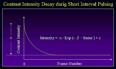
    The intensity gradually decreased to a constant value after switching from the long interval pulsing to short interval pulsing. The alteration in intensity is shown in Figure 1. The magnitude of intensity decay was greater for slower flow. An inverse correlation with flow velocity was also seen for the rate of intensity decay.


    Figure 1. Alteration in the contrast intensity decay during short interval pulsing in the in vitro study.

    PAGE TOP


    In vivo study


    An in vivo study using this method was then conducted in Sprague Dawley rats. The SONOS 5500 with a 12 MHz sector transducer in the acoustic densitometry mode was used to visualize left ventricular (LV) short axis images. Microbubbles were continuously infused through the femoral vein. Recordings with pulsing interval switching were performed at baseline and during pharmacological hyperemia.

    Results

    At baseline, the intensity from the anterior wall decreased more rapidly. During hyperemia the intensity was reduced slowly with a small extent of decay in total.

    PAGE TOP


    Proposed mechanism for the observation


    Oshita reviewed the mechanism for the observed relation between intensity decay and velocity. During a slow flow velocity, each pulse destroys a portion of the microbubbles in the imaging field. When the flow is slow, the bubble replenishment is slow, resulting in a rapid decrease in microbubble concentration. However, at a faster flow velocity, more time is required to attain the equilibrium between bubble destruction and influx. The equilibrium is reached at the lower level of bubble concentration for slow flow and the higher level for the faster flow, depending on the number of bubbles present in the imaging filed between pulses.

    PAGE TOP


    Report Index | Previous Report | Next Report
    Scientific Sessions | Activities | Publications
    Index

    Copyright © 2000 Japanese Circulation Society
    All Rights Reserved.

    webmaster@j-circ.or.jp