Japanese Circulation Society
Scientific Sessions Activities Publications
index
Congress Report
the 70th Scientific Session
Symposia
Symposium 7

Advances in Cardiovascular Imaging

Quantitative Assessment of Coronary Endothelial Function Using PET and Effects of Angiotensin II Receptor Blockade Therapy in Patients with Hypertension
Masanao Naya
Hokkaido University Graduate School of Medicine, Sapporo, Japan

Recent Advances in Intravascular Imaging Techniques
Shigeho Takarada
Wakayama Medical University, Wakayama, Japan


Quantitative Assessment of Coronary Endothelial Function Using PET and Effects of Angiotensin II Receptor Blockade Therapy in Patients with Hypertension

Masanao Naya

Hokkaido University Graduate School of Medicine
Sapporo, Japan


Dr. Masanao Naya, Hokkaido University, presented results of a study using positron emission tomography (PET) to evaluate the response of myocardial blood flow (MBF) to the cold pressor test (CPT) and the effects of angiotensin II receptor blockade (ARB) therapy on endothelial function.

PET with 15O-labeled water is a non-invasive method for obtaining quantitative sequential measurements of MBF. CPT uses sympathetic stimulation to induce endothelium dependent vasodilation via nitric oxide (NO) production. By measuring the change between MBF at rest and during CPT, the CPT-flow reserve (CPT-FR) can be calculated and used as a measure of endothelial function. Coronary vascular resistance (CVR) can be calculated by dividing the mean blood pressure (BP) by the MBF, to correct for the effects of BP.

The objectives of this study were to compare coronary endothelial function in hypertensive patients and healthy controls using 15O-labeled water PET; and to assess the effects of long-term ARB therapy on coronary endothelial function. The study included 10 hypertensive patients aged 20 to 70 years with mild to moderate untreated essential hypertension (systolic BP 146 ± 15 mmHg) without left ventricular hypertrophy (LVH) (LVMI 90 ± 21) and 10 control patients with systolic BP 110 ± 10 mmHg and no LVH (LVMI 85 ± 7).

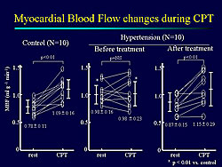
Figure 1. Myocardial blood flow was similar in the hypertensive patients and the control patients after treatment with olmesartan.
Click to enlarge

The hypertensive patients were treated with the ARB olmesartan (mean 27 ± 14 mg/day) for 12 weeks. MBF at rest and during CPT was measured by 15O-labeled water PET at baseline and after ARB treatment. Change between MBF at rest and during CPT, CPT-FR, and CVR were calculated.

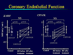
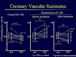
At baseline, compared to controls, the hypertensive patients had significantly higher systolic BP at rest (145 ± 14 versus 116 ± 14; p <0.01) and during CPT (161 ± 23 versus 133 ± 20; p <0.05). Prior to ARB treatment, MBF was significantly impaired both at rest and during CPT in the hypertensive patients compared to controls (p <0.01). After treatment, MBF of the hypertensive patients was similar to that of control patients (Figure 1). Coronary endothelial function, as measured by change in MBF and CPT-FR, was improved in hypertensive patients after ARB treatment compared to baseline (Figure 2). Before treatment, CVR was significantly higher in hypertensive patients than control patients during CPT (p <0.01). After ARB treatment, CVR during CPT was significantly decreased from baseline in the hypertensive patients (Figure 3).

Figure 2. Coronary endothelial function improved in the hypertensive patients with olmesartan treatment.
Click to enlarge

Figure 3. Coronary vascular resistance significantly improved with olmesartan treatment in the hypertensive patients.
Click to enlarge

Naya concluded that ARB therapy restores coronary endothelial function in hypertensive patients, as measured by change in MBF, CPT-FR, and CVR during CPT. PET with 15O-labeled water enables noninvasive quantitative estimation of MBF and is useful for monitoring the effects of medical therapy on coronary vasomotor function.


Recent Advances in Intravascular Imaging Techniques

Shigeho Takarada

Wakayama Medical University
Wakayama, Japan


Intravascular ultrasound (IVUS) is useful for evaluating the pathophysiology of atherosclerotic plaques in blood vessel walls. However, this method has limitations for assessing some tissue components in plaques. Dr. Shigeho Takarada, Wakayama Medical University, described studies of a new intravascular imaging technique, called optical coherence tomography (OCT), that might provide improved imaging of vulnerable plaques.

OCT is a new intravascular imaging method with a high resolution of 10 to 20 µm, which is 10-fold higher than the resolution of IVUS. OCT images show that fibrous plaques are signal rich and homogeneous with low attenuation, calcified plaques are signal poor and well-delineated with sharp borders, and lipid-rich plaques are signal-poor with diffuse borders.

While sensitivity of IVUS and OCT for diagnosing fibrous and fibrocalcific plaques is similar, sensitivity for lipid plaques is significantly higher with OCT (85%) than IVUS (59%) (p <0.05). OCT also has good correlation with histology for measuring thickness of fibrous caps. In a study of OCT to differentiate between red and white thrombi, peak intensity was similar for both types, but the intensity half distance was significantly higher for the white thrombus (324 ± 50) than the red thrombus (183 ± 42) (p=0.0001). OCT also allows quantification of macrophages in plaques.

Dr. Takarada’s group used an intra-coronary thermometer to identify areas in plaques with high numbers of macrophages. In a case study of a 64-year old female with unstable angina, use of the intra-coronary thermometer showed an 0.18°C difference in temperature between normal artery and the stenotic site. This technique was used to evaluate 34 lesions in 23 patients (14 males, 9 females) with stable effort angina and nine patients (7 males, 2 females) with unstable angina. Temperature of the intra-coronary artery was measured using an intra-coronary thermometer with a pressure guide wire, directional coronary atherectomy was performed, and the lesions were examined histologically with hematoxylin-eosin (H&E) stain, CD-68 for macrophages, and CD-45 for T cells.

Patients with unstable angina had significantly higher plaque temperatures (0.29 ± 0.16) than patients with stable angina (0.13 ± 0.10) (p=0.001). Patients whose plaques had high numbers of macrophages had higher temperatures (0.34 ± 0.15) than those whose plaques had low numbers of macrophages (0.11 ± 0.07) (p =0.0015).

Takarada concluded that recent advances in intravascular techniques, OCT, and temperature evaluation may enable identification of vulnerable plaques, including lipid-rich plaques, thin fibrous caps, thrombi, and high-macrophage plaques.


Copyright © 2006 Japanese Circulation Society
All Rights Reserved.
webmaster@j-circ.or.jp